Home
/ Mouth Anatomy Diagram : Human Mouth Anatomy In Detail - If yo wish you to know more about the anatomy of the mouth, look at the first diagram below.
Mouth Anatomy Diagram : Human Mouth Anatomy In Detail - If yo wish you to know more about the anatomy of the mouth, look at the first diagram below.
Mouth Anatomy Diagram : Human Mouth Anatomy In Detail - If yo wish you to know more about the anatomy of the mouth, look at the first diagram below.. The digestive tract (or gastrointestinal tract) is a long twisting tube that starts at the mouth and ends at the anus. The mouth (oral cavity) consists of several components, including the teeth, gingiva (gums), tongue, palate, cheeks, lips and floor of the mouth. Start studying full mouth anatomy diagram. Kidshealth offers a digital diagram with the basic parts of the mouth suited for kids at this age. The object of this game is to point and teach about each part of the mouth.
The oral cavity, better known as the mouth, is the start of the alimentary canal. With the exception of the teeth, the mouth is lined by mucous membranes. Focuses on the structures of the head and neck of the human body, including the brain, bones, muscles, blood vessels, nerves, glands The floor of mouth is an oral cavity subsite and is a common location of oral cavity squamous cell carcinoma. Related posts of human mouth anatomy diagram human ear anatomy diagrams.
Oral Cavity Anatomy Functions And Diseases Medical Library from d3uigcfkiiww0g.cloudfront.net This diagram depicts the human mouth.human anatomy diagrams show internal organs, cells, systems, conditions, symptoms and sickness information and/or tips for healthy living. The shape of your jaws also affects the way your face looks. The vestibule, the area between the cheeks and the teeth, and the oral cavity. Converging muscles around the mouth. Human mouth anatomy, structure, location and various functions. Mentalis, a small conical muscle originates from the incisive fossa of the mandible and is added into the skin of the lower lip. Focuses on the structures of the head and neck of the human body, including the brain, bones, muscles, blood vessels, nerves, glands Anatomy of the mouth ppt diagram take a closer look at each part of the mouth with the anatomy of the mouth ppt diagram.
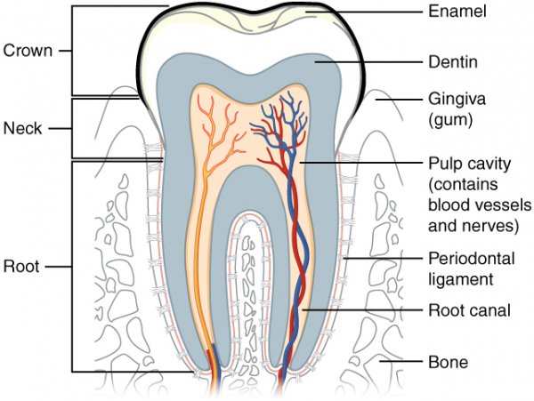
Teeth are the more characteristic part of the mouth's anatomy.
It puckers the chin and protrudes the lower lip. Let's start by examining the external anatomy of a fish. The mouth also plays a major role in the production of speech through the movements of the tongue, lips and cheeks. Twelve molars are usually present in an. This sheet helps you understand how the teeth and jaws work. The hyoid bones that connect the back of the tongue and the jaw are also connected to a group of muscles extending to the sternum, shoulder, and forelegs. It is found in most mammals that use their posterior teeth to grind food. Sublingual gland and its duct. Learn vocabulary, terms, and more with flashcards, games, and other study tools. Converging muscles around the mouth. But the components of the mouth and throat serve many functions. The anatomy of the mouth ppt diagram is a fully customizable powerpoint slide that will allow you to change the colors as you see fit. A fish's anatomy can be divided into external and internal.
Anatomy of the floor of the mouth the floor of the mouth is a horizontally aligned u shaped space situated in the part of the oral cavity that lies beneath the tongue. Based on anatomy, throat can be divided into 3 parts namely, the upper part, the middle part and the lower part called as nasopharynx, oropharynx and laryngopharynx respectively. The shape of your jaws also affects the way your face looks. With the exception of the teeth, the mouth is lined by mucous membranes. Start studying full mouth anatomy diagram.
34 1e Digestive System Mouth And Stomach Biology Libretexts from s3-us-west-2.amazonaws.com Looking at the diagram of the mouth above, the mouth structures includes the lips, cheeks, palate (roof of the mouth), floor of the mouth and the part of the tongue in the mouth (oral tongue). This diagram depicts the human mouth.human anatomy diagrams show internal organs, cells, systems, conditions, symptoms and sickness information and/or tips for healthy living. They are white colored calcified organs embedded partially in sockets of the lower and upper jaws. The major salivary glands, three pairs in total, are found in and around your mouth and throat. The mouth is the gate that opens so we can begin the process of energy absorption called digestion. It has three major functions: Sublingual gland and its duct. The nasopharynx is the uppermost part of the pharynx and extends down from the base of the skull to the nasal passages, hence the name.
The major salivary glands are the parotid, submandibular, and sublingual glands.the parotid glands are located in front and beneath the ear.
It is generally greatest in the incisor region (3.5 to 4.5 mm in the maxilla and 3.3 to 3.9 mm in the mandible) and less in the posterior segments, with the least width in the first premolar area (1.9 mm in the maxilla and 1.8 mm in the mandible). Mentalis, a small conical muscle originates from the incisive fossa of the mandible and is added into the skin of the lower lip. The external anatomy of a fish includes the fins, scales, gills, eyes, nares, mouth, lateral lines and vents. The shape of your jaws also affects the way your face looks. Point to a part and ask them how they think it might work. Basic mouth and throat anatomy for anesthesia and ent rotations Its main function is to help the horse eat and swallow food. A jaw that's too small, too large, or crooked can cause problems with chewing, speaking, breathing, and even sleeping. A fish's anatomy can be divided into external and internal. A molar tooth is located in the posterior (back) section of the mouth. Understanding jaw (orthognathic) anatomy and problems. A mucous membrane lines and protects the inside of the mouth. The vestibule, the area between the cheeks and the teeth, and the oral cavity.
Anatomy of the mouth ppt diagram take a closer look at each part of the mouth with the anatomy of the mouth ppt diagram. The mouth is the gate that opens so we can begin the process of energy absorption called digestion. It has three major functions: The vestibule, the area between the cheeks and the teeth, and the oral cavity. They are white colored calcified organs embedded partially in sockets of the lower and upper jaws.
Anatomy Human Mouth Sagittal Plane Soft Palate Tongue Text People Head Png Pngwing from w7.pngwing.com But the components of the mouth and throat serve many functions. Kidshealth offers a digital diagram with the basic parts of the mouth suited for kids at this age. The object of this game is to point and teach about each part of the mouth. This sheet helps you understand how the teeth and jaws work. It has three major functions: The floor of mouth is an oral cavity subsite and is a common location of oral cavity squamous cell carcinoma. It is made up of a series of muscles that coordinate the movement of food and. The tongue is made up of twelve highly sensitive muscles that react to pressure, pain, taste, and heat.
The hyoid bones that connect the back of the tongue and the jaw are also connected to a group of muscles extending to the sternum, shoulder, and forelegs.
Kidshealth offers a digital diagram with the basic parts of the mouth suited for kids at this age. With the exception of the teeth, the mouth is lined by mucous membranes. Let's start by examining the external anatomy of a fish. Sublingual gland and its duct. A mucous membrane lines and protects the inside of the mouth. Also known as the oral cavity, the mouth is the hollow cavity that allows food and air to enter the body. A duct, called stensen's duct, drains saliva from the parotid gland into the mouth, at the area of the upper cheeks. Converging muscles around the mouth. The teeth are held within the jaw bones and serve several important functions beyond allowing. This diagram depicts the human mouth.human anatomy diagrams show internal organs, cells, systems, conditions, symptoms and sickness information and/or tips for healthy living. Start studying full mouth anatomy diagram. Teeth are the more characteristic part of the mouth's anatomy. They are white colored calcified organs embedded partially in sockets of the lower and upper jaws.